


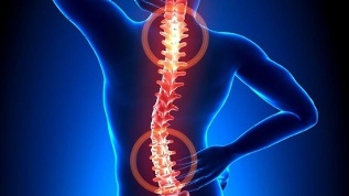
Excessive back muscle tension causes a lot of discomfort and pain. Osteochondrosis, which causes a violation of the structure of the vertebral and intervertebral discs, causes serious pinching of nerve endings. Often, the pathology is accompanied by deterioration of blood circulation, which causes disturbances in the nutrition of the brain and internal organs.
Osteochondrosis - what is it?

Osteochondrosis is a recurrent disease that occurs in a chronic form and is accompanied by destruction of the vertebrae with the intervertebral disc. Their tissues are disturbed, which provokes a decrease in their level of elasticity, followed by deformation. There is a gradual decrease in the intervertebral space. This causes loss of spinal space stability in areas of pathological development.
The process of destruction of pathological tissue occurs against the background of the pinched nerve endings, which are directed from the area where the spinal cord is located. As a result, the back muscles are in a constant state of tension. In such cases, the patient complains of back pain and other symptoms.
Based on the uniqueness of localization of spinal structure, which is covered by degenerative changes, the types of pathological processes of the cervix, thorax and lumbosacral are distinguished. The main symptoms of the development of osteochondrosis are pain, intensity and severity that usually increase during physical exercise.
There is also stiffness in movement. In addition, the clinical picture is characterized by the presence of signs of vertebral type - headache, changes in blood pressure, deterioration of visual function, hearing, and so on.
Development Mechanism
The development of osteochondrosis is associated with the fact that the nucleus pulposus begins to lose its hydrophilic quality. This semi-liquid structure contains connective tissue fibers and chondroitin, a gelatinous material. In the process of expansion of the human body and its growth, the process of reduction of the vascular bed on the intervertebral disc is actively underway. Nutrients are supplied in a permeable manner, which manifests itself in the stabilization of spontaneous concentrations. This feature is the reason for the difficulty in fully recovering cartilage that causes excessive damage or stress to the spine.
Pathological abnormalities become more pronounced due to violations of hormonal background and human nutrition. Cartilage tissue begins to lack nutrients necessary for its normal development. Thus, the disorder appears in the form:
- decreased strength and elasticity;
- changes to consistency parameters and configuration properties.
Against the background of leveling the intervertebral disc, the formation of radial fractures on the fibrous ring occurs. As a result, the intervertebral distance is reduced, and the facet joints begin to shift. Over time, pathological changes include types of connective tissue associated with fibrous rings and ligaments.
When tissue is broken down by the immune system, an increase in the amount of immunoglobulins is produced. This provokes the development of aseptic inflammatory processes, edema forms in the area where the facet joint is located. They also spread to nearby soft tissues.
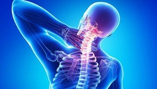
Due to the stretching of the joint capsule, the intervertebral disc loses the ability to repair the vertebrae. The instability of the position of such spinal structures increases the risk of pinching nerve roots or squeezing blood vessels. This feature is typical, for example, for cervical osteochondrosis, which is accompanied by strong oral symptoms.
Causes of disease
The condition of the intervertebral disc can worsen with decreased skeletal muscle tone in the spine. Due to irrational and asymmetric muscle work, destruction of cartilage tissue may occur with prolonged maintenance of non-physiological positions of the body. This violation is the result of wearing a heavy bag on the same shoulder, using a soft mattress and high pillows.
The process of destruction of the intervertebral disc is accelerated due to the action of a number of negative factors of external and internal nature. These include:
- disorders of endocrine mechanisms and metabolic disorders;
- pathology is contagious, including in chronic form;
- injury to spinal space in the form of compression fractures, bruises;
- normal and prolonged hypothermia of the body;
- systemic and degenerative-dystrophic diseases - gout, psoriatic, rheumatoid arthritis, osteoporosis, osteoarthritis;
- smoking and alcohol abuse, which disrupt the state of the vascular system, disrupt blood circulation and provoke nutrient deficiencies in cartilage;
- inadequate physical development, problems with posture, flat feet - these deformities increase the load on the spinal space, as amortization will not be sufficient;
- obesity;
- genetic predisposition;
- exposure to normal stress.
Symptoms

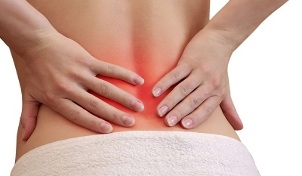
The main clinical sign of osteochondrosis of any localization (cervical, thoracic or lumbosacral) is pain syndrome. With relapse, the pain penetrates, radiating to nearby body areas. Even with a little movement, it is growing. This forces the patient to place the torso in a forced position to reduce discomfort and pain:
- with cervical osteochondrosis, it is better to turn one head, but the whole body;
- when there is a form of pain in the chest, it is difficult for the patient to breathe deeply, and therefore, to exclude acute chest pain, he seeks to reduce the depth and frequency of breathing;
- in patients with this type of lumbar disease, difficulty arises when they sit, take an upright position, move, when the nerve location of the spine is pinched.
Typically, patients complain of dull, persistent pain and cramps in movement in the morning after waking up. In this case, a differential diagnosis is needed to help eliminate the risk of developing myositis caused by inflammation of the skeletal muscle or osteoarthritis.
Pain and stress that arise due to compensatory tension in muscle tissue. This condition is necessary to stabilize the area of movement of the spine. Persistent mild to moderate pain may appear with significant intervertebral disc stretching, and is caused by aseptic inflammatory changes.
Separate localized osteochondrosis is characterized by special symptoms:
- With cervical osteochondrosis, pain is felt in the cervical zone, in the upper limbs. Headache and numbness of the fingers were observed. If the disease manifests itself in serious form, then pinching of the vertebral arteries can occur. In this case, the patient begins to complain of significant health deterioration.
- Localization of the thorax is indicated by acute pain and pain in the back, visceral pain syndrome present in the heart, right hypochondrium and abdomen. Patients complain of numbness, paresthesia on the skin, shortness of breath, cramps in the vertebrae.
- Patients with lumbar osteochondrosis complain of pain in the back and lower legs with increased intensity when moving. Often, disorders in the organ function of the genitourinary system, problems with male potency, dysfunctional ovarian disorders are diagnosed. During remission, the pain can be reduced. However, the effects of the provoking factors led to its reform.
- When mixed osteochondrosis manifests itself, symptomatology can manifest itself in several zones at the same time. This condition is characterized by more severe disease.
Keep in mind that vertebral displacement and osteophyte formation cause compression of the vertebral arteries. It nourishes the brain, providing its cells with oxygen components. When squeezed, food is limited, and therefore patients have problems with coordination, headaches, tinnitus, and arterial hypertension.
Consequences if not treated
The cause of complicated osteochondrosis is the rapid formation of hernias in the intervertebral disc. Their appearance is associated with a displacement of the vertebral structure toward the posterior. This triggers the rupture of the posterior ligament of the longitudinal type, resulting in instability of the disc position, protrusion of individual parts into the spinal canal area. The rupture of a hernia occurs when a disc with the nucleus pulposus penetrates into the duct area.
With the manifestation of pathological abnormalities in the structure of the vertebrae, the back of the brain begins to squeeze, the patient develops discogenic myelopathy. Symptoms of this condition are associated with numbness and weakness in certain muscle groups in the upper and lower extremities. Paresis, muscle atrophy, and tendon reflexes are indicated. In some cases, there is a problem with emptying the bladder, with the intestines.
Disc herniation is dangerous by pressing on the arteries that supply the spinal cord. The result of this pathology is the formation of ischemic zones, where nerve cells have suffered damage and death. Manifestations of neurological effects are expressed in motor function failure, decreased tactile levels, and trophic disorders.
Disease Diagnostics
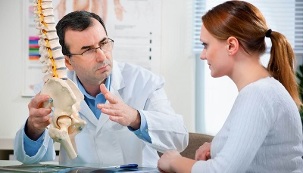
The initial diagnosis is made based on the patient's complaints and symptoms. Specialists study the condition of the spinal space in different positions, indicating the patient is at rest or moving. At the next stage, the patient is sent for laboratory diagnostics, which will help clarify the diagnosis or refute it.
The research methods used include:
- Radiography- provides a complete examination of the spinal space with an assessment of the condition of the spine, disorders in the form of growth, curvature. The specialist will be able to determine the intervertebral type interval, the condition of the hole. To accurately identify osteochondrosis, which is localized in the chest or cervical area, a two-stage x-ray examination is performed. In the first stage, the patient lies on his side, and in the second stage, right on his back.
- The tomography method using MRI or CTprovides very informative data, which helps to study the vertebrae in detail without interruption in the shape of the organ that covers it. The picture shows the nerves and the vascular system. MRI helps identify the signs of many diseases of the spine and the location of the damage. With CT, the hernia is visualized, the possibility of abnormalities in the spinal structure is determined.
- Laboratory examinationto assess blood condition and its main parameters. Allows you to clarify the diagnosis and determine the likelihood of having the same disease.
In many cases, as a result of the examination, the doctor diagnoses the presence of several background diseases, which potentially endanger their complications. We talk, for example, about hernias, protrusions, radiculitis. Proper problem diagnosis helps treat osteochondrosis effectively. In this case, the disease itself in the early stages of its development disguises itself as a symptom of another disease.
Therapy Process
Osteochondrosis is treated conservatively or surgically. The choice depends on the severity of the condition, its neglect, the degree of tissue deterioration, and its cause.
It is important to remember that it is impossible to completely cure osteochondrosis, as there is no cure to help restore the disc and vertebrae completely. The therapeutic effect is focused on inhibiting the process of destruction and increasing the duration and stability of remission.
For symptomatic therapy, chondroprotectors are used, which are based on chondroitin sulfate or glucosamine.
The effectiveness of the therapeutic process with the use of chondroprotectors has been clinically confirmed by long-term trials. If you take this fund for a long period of 3 months, then there is some recovery of cartilage and other elements of the connective type - ligament-tendon apparatus, exchange.
Accumulation of glucosamine and chondroitin in the intervertebral disc area leads to manifestations of analgesic, anti-edematous and anti-inflammatory effects. Therefore, there is a real opportunity to optimize the dose of NSAIDs, glucocorticosteroid group drugs, muscle relaxants. You can rely on a reduction in the burden of medication on the patient.
The effectiveness of chondroprotectors is determined by the regularity of its intake. Otherwise, there will be no results. Ineffectiveness was also noted in the treatment of osteochondrosis at grade 3, accompanied by significant cartilage destruction.
The following groups of drugs can be used to relieve pain:
- Nonsteroidal anti-inflammatory drugshelp eliminate inflammatory disorders in the soft tissues caused by vertebral transplants. NSAIDs are effective in reducing pain, swelling, and stiffness.
- Facilities of the glucocorticosteroid group- usually a restriction used in conjunction with anesthesia. They can relieve pain, restore immune mechanisms, and provide anti-exudative effects.
- Relax the muscles.They are effective in combating muscle cramps due to nerve entrapment. They help loosen skeletal muscle and block polysynaptic spinal reflexes with antispasmodic effects.
- External medicine with heating effect.Irritation of subcutaneous tissue receptors with activation of blood flow is provided by special gels and ointments. This drug has analgesic and anti-edematous effects.
It is possible to get rid of the symptoms of the vertebrogenic type, which manifests itself as a result of pathological localization in the cervical or thoracic zone, using medical devices to activate blood flow. Nootropics and drugs to improve microcirculation are also prescribed. In some cases, you may need to take antidepressants, as well as medications with anticonvulsants.
During the treatment of osteochondrosis, physical therapy is also used. UHF therapy procedures, magnetotherapy, laser therapy, reflexology, massage, exercise therapy, hirudotherapy, as well as swimming and yoga can be prescribed. If conservative treatment is ineffective, surgery is performed using microdiscectomy, puncture valorization, laser reshaping, or implant replacement.